How do cells build and maintain compartments?
Membranes are the bags that hold cells together. Within eukaryotic cells, sub-compartments are formed by organelle membranes to allow specialized structures. Despite the essential nature of membranes to eukaryotic life, there are still many questions that remain unanswered:
- How are a diverse array of organelle membranes constructed and maintained by the cell?
- What is the starting material for the biogenesis of a particular membrane?
- How is the biogenesis of membranes coordinated with the cell cycle?
Our lab tackles these questions by studying the model organism, S. pombe.
Meiosis II in S. pombe
Our approaches
Live-cell fluorescence microscopy
We use live-cell imaging to capture dynamic events such as mitosis and meiosis. Here, you can see S. pombe undergoing mitosis with labelled nuclear envelope (green) and microtubules (magenta) over the course of 30 min.
Correlative light and electron microscopy (CLEM)
When we need to directly visualize membranes, we use electron tomography (ET). Often, we’ll use fluorescence imaging to inform our electron tomography, as was the case in finding this cell in late anaphase B. Here, the mitotic nuclear envelope (blue), microtubules (yellow), and spindle pole body (orange) are annotated.
Yeast genetics
Fission yeast are a fantastic model organism to examine the genetic interactions. By comparing the size of colonies on this plate, we were able to demonstrate a synthetic sick interaction between the CMP7 and PCP1 genes. This informed our further functional analysis by microscopy.
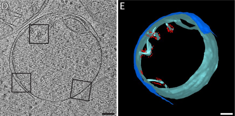
Electron Cryo-Tomography (cryo-ET)
Sometimes, we want to see more than just membranes by electron microscopy. In that case, we turn to electron cryo-tomography (cryo-ET) mediated by cryo-focused ion beam milling. Shown here are the ATP synthases (red) mislocalizing during Bax-induced apoptosis in the mitochondrion (blues) of a HeLa cell.